


 Foundational characteristics of cancer include proliferation, angiogenesis, migration, evasion of apoptosis, and cellular immortality. Find key markers for these cellular processes and antibodies to detect them.
 The SUMOplot™ Analysis Program predicts and scores sumoylation sites in your protein. SUMOylation is a post-translational modification involved in various cellular processes, such as nuclear-cytosolic transport, transcriptional regulation, apoptosis, protein stability, response to stress, and progression through the cell cycle.
 The Autophagy Receptor Motif Plotter predicts and scores autophagy receptor binding sites in your protein. Identifying proteins connected to this pathway is critical to understanding the role of autophagy in physiological as well as pathological processes such as development, differentiation, neurodegenerative diseases, stress, infection, and cancer.
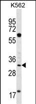
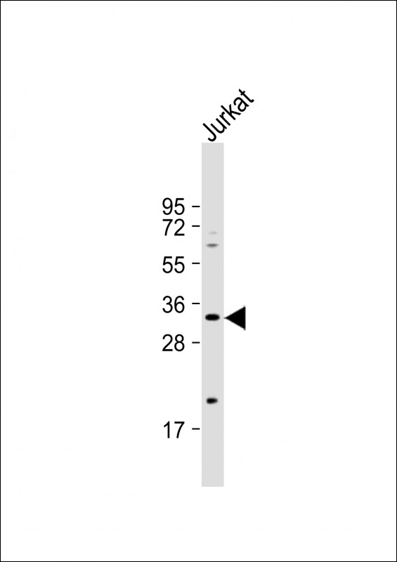
FCN3 Antibody (C-term)
Affinity Purified Rabbit Polyclonal Antibody (Pab)
- SPECIFICATION
- CITATIONS
- PROTOCOLS
- BACKGROUND

Application
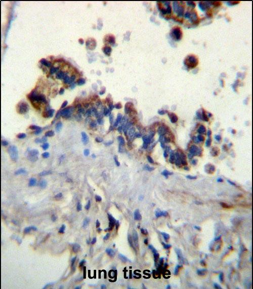
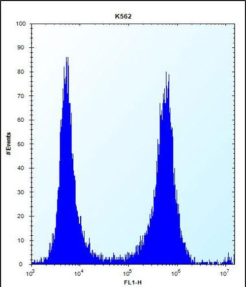
| WB, FC, IHC-P, E |
|---|---|
| Primary Accession | O75636 |
| Other Accession | NP_775628.1, NP_003656.2 |
| Reactivity | Human |
| Host | Rabbit |
| Clonality | Polyclonal |
| Isotype | Rabbit IgG |
| Calculated MW | 32903 Da |
| Antigen Region | 214-243 aa |
| Gene ID | 8547 |
|---|---|
| Other Names | Ficolin-3, Collagen/fibrinogen domain-containing lectin 3 p35, Collagen/fibrinogen domain-containing protein 3, Hakata antigen, FCN3, FCNH, HAKA1 |
| Target/Specificity | This FCN3 antibody is generated from rabbits immunized with a KLH conjugated synthetic peptide between 214-243 amino acids from the C-terminal region of human FCN3. |
| Dilution | WB~~1:500 FC~~1:10~50 IHC-P~~1:10~50 E~~Use at an assay dependent concentration. |
| Format | Purified polyclonal antibody supplied in PBS with 0.09% (W/V) sodium azide. This antibody is purified through a protein A column, followed by peptide affinity purification. |
| Storage | Maintain refrigerated at 2-8°C for up to 2 weeks. For long term storage store at -20°C in small aliquots to prevent freeze-thaw cycles. |
| Precautions | FCN3 Antibody (C-term) is for research use only and not for use in diagnostic or therapeutic procedures. |
| Name | FCN3 |
|---|---|
| Synonyms | FCNH, HAKA1 |
| Function | May function in innate immunity through activation of the lectin complement pathway. Calcium-dependent and GlcNAc-binding lectin. Has affinity with GalNAc, GlcNAc, D-fucose, as mono/oligosaccharide and lipopolysaccharides from S.typhimurium and S.minnesota. |
| Cellular Location | Secreted. Note=Found in blood plasma, bronchus, alveolus and bile duct |
| Tissue Location | Liver and lung. In liver it is produced by bile duct epithelial cells and hepatocytes. In lung it is produced by both ciliated bronchial epithelial cells and type II alveolar epithelial cells. |

Thousands of laboratories across the world have published research that depended on the performance of antibodies from Abcepta to advance their research. Check out links to articles that cite our products in major peer-reviewed journals, organized by research category.
info@abcepta.com, and receive a free "I Love Antibodies" mug.

Provided below are standard protocols that you may find useful for product applications.
Background
Ficolins are a group of proteins which consist of a collagen-like domain and a fibrinogen-like domain. In human serum, there are two types of ficolins, both of which have lectin activity. The protein encoded by this gene is a thermolabile beta-2-macroglycoprotein found in all human serum and is a member of the ficolin/opsonin p35 lectin family. The protein, which was initially identified based on its reactivity with sera from patients with systemic lupus erythematosus, has been shown to have a calcium-independent lectin activity. The protein can activate the complement pathway in association with MASPs and sMAP, thereby aiding in host defense through the activation of the lectin pathway. Alternative splicing occurs at this locus and two variants, each encoding a distinct isoform, have been identified.
References
Davila, S., et al. Genes Immun. 11(3):232-238(2010)
Andersen, T., et al. J. Rheumatol. 36(4):757-759(2009)
Ruskamp, J.M., et al. Clin. Exp. Immunol. 155(3):433-440(2009)
Lacroix, M., et al. J. Immunol. 182(1):456-465(2009)
Munthe-Fog, L., et al. Mol. Immunol. 45(9):2660-2666(2008)
If you have used an Abcepta product and would like to share how it has performed, please click on the "Submit Review" button and provide the requested information. Our staff will examine and post your review and contact you if needed.
If you have any additional inquiries please email technical services at tech@abcepta.com.
Ordering Information
Other Products
Shipping Information
